Gall bladder
GALL BLADDER
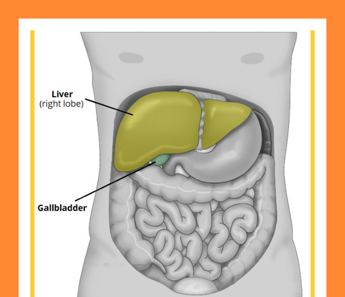


The gallbladder is a digestive organ that is situated in the right hypochondrium of the abdomen. This pear-shaped intraperitoneal sac is located in a fossa that was created by the inferior aspects of the right and quadrate lobes of the liver.
Concentrating and storing bile, which is created by the liver, is the gallbladder's main job. The stored bile is then released from the gallbladder as part of the gustatory response in response to cholecystokinin.
In this article, we'll examine the gallbladder's anatomy, including its structure, vascular, innervation, and lymphatic supply.
Anatomical Relations
The visceral surface of the liver is directly above the gallbladder, which is completely enclosed by peritoneum.
The following buildings are situated nearby:
The inferior hepatic border and the anterior abdominal wall are located anteriorly and superiorly.
The proximal duodenum and the transverse colon are located posteriorly.
The remaining duodenal segments and the biliary tree are located inferiorly.
Anatomical Structure
The gallbladder resembles a pear-shaped cul-de-sac and connects to the common hepatic ducts via the cystic duct. The sac actually appears grey-blue in life (and not green as depicted in the texts). The 7.5 to 12 cm long organ is located deep to the hepatic section of the peritoneum, on the inferior face of the anatomical right lobe of the liver.
The first segment of the duodenum is located anterior to the gallbladder, which has a storage capacity of 30 to 50 ml. Typically, it is broken into three sections:
The fundus is the spherical, distal part of the gallbladder. It extends into the liver's inferior surface in the mid-clavicular line.
The body of the gallbladder is its major component. It is situated next to the transverse colon, superior portion of the duodenum, and the posteroinferior aspect of the liver.
Neck: The gallbladder tapers and merges with the cystic duct at this point, entering the biliary tree.
.. A mucosal fold called Hartmann's Pouch can be found on the neck. Cholestasis is frequently brought on by gallstones that have been trapped in this area.
The Biliary Tree.
The biliary tree is a network of gastrointestinal ducts that allows the liver to concentrate and store newly synthesised bile in the gallbladder (prior to release into the duodenum).
The left and right hepatic ducts receive the bile that is first released by hepatocytes and then drains from both liver lobes via canaliculi, intralobular ducts, and collecting ducts. Together with the hepatic vein, these ducts combine to form the common hepatic duct.
The cystic duct joins the common hepatic duct as it descends, allowing bile to enter and exit the gallbladder for storage and release. The common bile duct is now created by the union of the cystic and common hepatic ducts.
The initial section of the duodenum and the head of the pancreas are where the common bile duct descends and continues posteriorly. The hepatopancreatic ampulla, also called the ampulla of Vater, is formed at this location when the main pancreatic duct joins it. The ampulla of Vater then empties into the duodenum via the major duodenal papilla. The sphincter of Oddi, a muscle valve, controls this papilla.
Vasculature
The cystic artery, a branch of the right hepatic artery, provides blood flow to the gallbladder (which itself is derived from the common hepatic artery, one of the three major branches of the coeliac trunk).
The cystic veins that drain directly into the portal vein are used for venous drainage of the gallbladder's neck. The hepatic sinusoids receive the venous outflow from the gallbladder's fundus and body.
Innervation
The gallbladder is sensory, sympathetic, and parasympathetically innervated.
Parasympathetic innervation is delivered by the vagus nerve, whereas sympathetic and sensory fibres are carried via the CELIAC PLEXUS.
Due to the relaxation of the sphincter of Oddi, parasympathetic activation causes the gallbladder to contract and the release of bile into the cystic duct. Nevertheless, circulating cholecystokinin, which is a component of the gustatory response, mediates the majority of this response.
Lymph Drainage
The cystic lymph nodes, which are located at the gallbladder neck, receive gallbladder-derived lymph.
The hepatic lymph nodes and finally the celiac lymph nodes are where the cystic nodes eventually empty.
Clinical Relevance: Gallstones//
Gallstones, or cholelithiasis, are tiny cholesterol, bile salt, or a combination of the two, lumps that can develop inside the gallbladder. They are typically asymptomatic and rather common.
But, they could also cause discomfort, a rash, and systemic ill health (depending on the location of the gallstone, and the presence or absence of associated infection or inflammation).
To differentiate between these diseases, various terms are used:
Uncomplicated gallstones are called cholelithiasis.
Biliary colic is characterised by pain in the right upper quadrant that usually develops after a fatty meal because gallstones block the cystic duct when the gallbladder contracts. not connected to a systemic disturbance.
Gallbladder inflammation is known as cholecystitis. Pain frequently coexists with sickness, vomiting, or fever.
Common bile duct gallstones are known as choledocholithiasis. Often results in abnormal liver function tests.
Infection of the common bile duct caused by choledocholithiasis is known as cholangitis. Right upper quadrant pain, fever, and jaundice are typical symptoms of Charcot's triad.
Most symptomatic individuals undergo surgical removal of the gallbladder (cholecystectomy) once they have been diagnosed; this procedure is now frequently carried out by laparoscopic (key-hole) surgery during the acute phase or after healing has taken place (often at 6 weeks).
Patients are given analgesics and antibiotics as needed in the interim.
Thank you ☺️
Best of luck 👍
zeeshanfayazlone@


Comments
Post a Comment